Doggy Dolly F018 Smoking für Hunde mit Fliege, grau/blau B00BD81OOO B00BD81OOO
- Handwäsche
- Hochzeitsmode für Hunde
- Klettverschluss am Bauch
- Dekoknöpfe an Weste und Hemd
- Rassenbeispiele Größe L: Shi Tzu, Pekinese, Miniatur Schnauzer, Pudel
Produktbeschreibungen
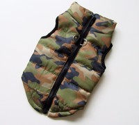
Größe: L Rückenlänge: 31-33cm; 12-13inch Brustumfang: 46-48cm; 18-19inch. Der blau-graue Smoking für den Hund ist ein echter Hingucker bei jeder Hochzeit - egal ob bei der Hundehochzeit oder wenn Herrchen Frauchen heiratet. Der Hundesmoking besteht aus einer kurzärmeligen Jacke und einem eingenähten Hemd. Natürlich dürfen auch Fliege und Krawatte bei diesem festlichen Outfit nicht fehlen. Fliege und Dekoknöpfe befinden sich bei diesem Modell auf dem Rücken. Die Weste ist für den Hund durch den Klettverschluss am Bauch einfach an- und auszuziehen. Der Anzug für Hunde hat Dekoknöpfe an Hemd und Weste. Doggydolly Hundemode gehört zu den weltweit führenden Herstellern im Bereich der Hundebekleidung und Accessoires für Hunde. Die Kleidungsstücke zeichnen sich durch ihre hochwertige Qualität sowie detailgenaue Verarbeitung aus. Doggydolly bietet eine große Auswahl im Bereich Brautmode und Festtagsmode für den Hund.
Stuhlfarbe
Die Farbe des Stuhls ist von den in der Galle enthaltenen Gallenfarbstoffen (Bilirubin und Biliverdin) abhängig.
Normalerweise ist die Stuhlfarbe hell- oder dunkelbraun, je nach Zusammensetzung:
- Bakterien,
- Wasser,
- Galle,
- Bilirubin,
- Hämoglobin,
- pflanzliche unverdauliche Substanzen, wie Zellstoff, Eiweiß und Fett.
Die Farbe des Stuhlgangs ist sehr aufschlussreich und kann auf wichtige Probleme hinweisen; anomale Farben sind:
Die Galle ist eine goldgelbe Flüssigkeit, die aus vielen chemischen Substanzen besteht, dazu gehört auch Bilirubin.
Die Galle wird von der Gallenblase gebildet und dann in den Darm befördert.
Hämoglobin ist ein in den roten Blutkörperchen enthaltenes Protein, das dem Sauerstofftransport dient.
Wenn die roten Blutkörperchen absterben, werden sie auf natürlichem Weg in Leber und Milz abgebaut.
Dabei wird das Hämoglobin freigesetzt.
Das Häm entsteht bei der Spaltung des Hämoglobins; es ist der Hauptbestandteil der Galle und gibt ihr ihre Farbe.
Wenn die Galle durch den Darm wandert, erlebt sie weitere chemische Veränderungen und verändert die Farbe.
Das in der Galle enthaltene Bilirubin degradiert im Darm zu:
- Urobilin, eine gelbliche Substanz, die über den Urin ausgeschieden wird,
- Stercobilin, eine bräunliche Mischung, die den Körper über den Kot verlässt.
Die Farbe von Stuhl und Urin werden durch diese beiden Substanzen bestimmt.
Wenn der Darm beispielsweise sehr schnell durchlaufen wird, hat die Galle wohlmöglich nicht genug Zeit, die Farbe zu ändern und der Stuhl hat eine grüne Färbung.
Die Farbe des Stuhls kann sich auch aus verschiedenen Gründen verändern.
- Eine Stuhlverfärbung kann harmlose Ursachen haben, besonders wenn sie nur einmal auftritt.
- Lang anhaltende Farbveränderungen des Stuhls können auf eine Krankheit hindeuten.
Roter Stuhl
Eine leuchtend rote Stuhlfärbung weisen auf Blutungen im unteren Darmabschnitt (Anus und Rektum) hin, die auftrund von Krankheiten wie Hämorrhoiden , Divertikulose oder bei schweren Erkrankungen wie Darmkrebs auftreten.
Rot (oder rötlich) gefärbter Stuhlgang kann aber auch durch den Verzehr von roter Lebensmittelfarbe oder roten Nahrungsmitteln wie Rote Bete und Tomatensaft verursacht werden.
Der Arzt sollte stets darüber informiert werden, auch wenn nicht immer eine ernsthafte Krankheit dahintersteckt.
Ursachen für roten Stuhl
Zur Behandlung von Hämorrhoiden und Analrhagaden sollte die Ernährung umgestellt werden:
- Mehr Obst, Nussfrüchte (ohne zu übertreiben) und Gemüse essen.
- Getreide einschränken; sie können durch Kartoffeln und Hülsenfrüchte ersetzt werden.
- Umgewandelte und frittierte Lebensmittel sowie Junkfood vermeiden.
- Einen Arzt konsultieren, um eine Blutung durch Krebs auszuschließen.
Dunkelroter oder bordeauxroter Stuhl
Eine dunkelrote Stuhlfarbe weist auf eine Blutung im Kolon oder Krankheiten im oberen Abschnitt des Verdauungsapparates hin.
Ursachen für dunkelroten Stuhl
- Darmparasiten oder Infektionen
- Divertikulitis
- Reizdarmsyndrom
- Tumoren im oberen Magen-Darm-Trakt
- Polypen
- Geschwüre
- Krankheiten der Speiseröhre
- Zu viel Alkohol
- Bestimmte Medikamente
- Verzicht auf Alkohol.
- Magenreizende Medikamente absetzen.
- Behandlung der Infektionen bzw. Ausrottung der Parasiten.
- Arztbesuch, um Blutungen des oberen Magen-Darm-Traktes auszuschließen.
Grüner Stuhl
Wenn der Darminhalt zu schnell in Richtung Rektum wandert, kann das in der Galle enthaltene grünfarbene Biliverdin auf dem Weg durch den Darm nicht schnell genug in seine braunen Abbauprodukte umwandeln.
Eine hellgrüne Stuhlfarbe kommt sehr häufig vor, auch nach dem Verzehr von Lebensmitteln, die Blattgrün (Chlorophyll) oder grüne Farbstoffe enthalten (wie Salat, Gartenrauke, Mangold, Spinat usw.).
Die hellgründe Färbung ist kein Hinweis auf eine Erkrankung.
Ursachen für hell- und dunkelgrünen Stuhl
- Durchfall
- Colitis ulcerosa
- Reizdarmsyndrom
- Verzehr von grünen Nahrungsmitteln
- Infektionen, z.B. durch Salmonellen
- Medikamente
- Arztbesuch, um Erkrankungen des Kolons auszuschließen.
Gelber Stuhl
Gelber, fetter und übel riechender Stuhl kann folgende Ursachen haben:
- Unfähigkeit, Fette zu verdauen aufgrund von Krankheiten, die die Darmschleimhaut angreifen, wie Zöliakie oder zystische Fibrose.
- Erkrankungen der Bauchspeicheldrüse, die die Produktion der Verdauungsenzyme vermindern, wie Pankreatitis.
- Verstopfung des Pankreasgangs, beispielsweise im Falle eines Tumors der Bauchspeicheldrüse.
- Unzureichende Gallenproduktion, zum Beispiel durch Verstopfung der Gallengänge oder Leberkrebs.
- Infektion und Vergiftung – Eine Infektion durch Giardia lamblia ruft einen typisch gelben Durchfall hervor. Die verschiedenen durch Viren, Bakterien und Parasiten ausgelösten Infektionen des Magen-Darm-Traktes können neben Durchfall auch eine Verfärbung des Stuhls verursachen.
Ursachen für gelben Stuhl
- Malabsorption der Fette
- Parasiteninfektionen
- Virusinfektionen, z.B. durch Rotavirus
- Bauchspeicheldrüsenkrebs
- Morbus Meulengracht
- Reizdarmsyndrom
- Zöliakie
- Verzehr von fetten Nahrungsmitteln
- Umstellung der Ernährung und Erhöhung der Ballaststoffzufuhr.
- Arztbesuch, um Erkrankungen der Leber und Bauchspeicheldrüse auszuschließen.
Weißer oder heller Stuhl
Ein weißgefärbter Stuhl kann durch ein Problem im Gallensystem entstehen, das sich wie folgt zusammensetzt:
- Bauchspeicheldrüse (Pankreatitis oder Pankreaskrebs)
- Leber (sklerosierende Cholangitis, Hepatitis, Leberzirrhose und Leberkrebs)
- Gallenblase (Gallensteine oder Gallenblasenentzündung)
- Das bei Röntgenuntersuchungen eingesetzte Barium kann dasselbe Aussehen bewirken.
- Schleim im Stuhl kann diesem eine weißliche Färbung verleihen und durch Entzündungen oder gutartige Erkrankungen, wie das Reizdarmsyndrom, entstehen.
- Erkrankungen wie: Pfeiffersches Drüsenfieber, zystische Fibrose und Sichelzellenanämie.
- Zu hoher Anteil von Reis und Kartoffeln in der Ernährung.
- Medikamente basierend auf Aluminiumhydroxid.
- Gesündere Ernährung.
- Depuration von Kolon und Leber.
Schwrazer Stuhl
Schwarzer Stuhl (Teerstuhl oder Meläna) können aufgrund von Blutungen im oberen Magen-Darm-Trakt (Speiseröhre, Magen, Dünndarm) auftreten, die schwarze Farbe bedeutet das Vorhandsein von verdautem Blut.
Dunkelgrauer Stuhl kann durch eine erhöhte Eisenkonzentration hervorgerufen werden, bedingt durch:
- Eisenpräparaten,
- den Verzehr von viel Fleisch oder Schokolade,
- wismuthaltigen Lebensmitteln.
n für Verzehr von dunklen Lebensmitteln (Aktivkohle, Lakritz, schwarze Oliven)
- Erhöhte Eisenzufuhr
- Alkoholmissbrauch
- Magen- oder Zwölffingerdarmgeschwüre
- Blutung durch Krampfadern der Speiseröhre (Ösophagusvarizen)
- Andere Blutungen im Magen-Darm-Trakt
- Gewisse Medikamente
- Bauchschmerzen
- Erbrechen
- Durchfall
- Schwäche
- Schwindel
- Bei verdautem Blut: sauer, fauliger, ekelerregender Geruch des Stuhls
- Verzicht auf Alkohol.
- Einnahme von Eisen vermindern.
- Auf magenreizende Medikamente verzichten.
- Arztbesuch, um Störungen des oberen Magen-Darm-Traktes auszuschließen.
Oranger Stuhl
In der Regel wird oranger Stuhl nicht durch eine Krankheit verursacht, sondern durch die Einnahme von Medikamenten oder den Verzehr von Lebensmitteln mit einem hohen Anteil an Betakarotin.
Ursachen für orangen Stuhl
- Verzehr von orangen Lebensmitteln (Möhre, Mango, Aprikose, Kürbis)
- Ergänzungsmittel mit Betakarotin
- Medikamente (z.B. Rifampicin)
Wann ist eine Stuhlverfärbung Grund zur Sorge?

Folgende Stuhlfarben sind untypisch und sollten unverzüglich ärztlich abgeklärt werden:
- Leuchtendes Rot
- Dunkelrot
- Schwarzer Teerstuhl
- Lehmfarbig oder weiß – hell
Stuhlfarbe bei Neugeborenen
Die Stuhlfarbe bei Neugeborenen ist abhängig von:
- Alter
- Milchtyp (Muttermilch oder Fertigmilch)
- Zugabe fester Beikost.
In den ersten beiden Tagen nach der Geburt scheidet das Neugeborene Kindspech oder Mekonium aus, das sich aus Schleim, Fruchtwasser und allem, was das Kind während der Zeit in der Gebärmutter verschluckt hat, zusammensetzt.
Mekonium hat eine schwärzlich-grüne Farbe und ist eine zähe klebrige Masse, wie Teer.
Das mitunter schwierig zu säubernde Kindspech ist ein Zeichen dafür, dass die Verdauung des Kindes normal funktioniert.
Wie ist der Neugeborenenstuhl beim Stillen?
Die Vormilch (Kolostrum) ist die erste Milch, die von der Milchdrüse produziert wird und hat eine abführende Wirkung, was die Ausscheidung des Kindspechs begünstigt.
Nach etwa drei Tagen wird die normale Milch erzeugt und der Stuhl ändert allmählich sein Aussehen.
In den ersten Tagen lässt er sich wie folgt beschreiben:
- Die Farbe ist hell, von gründlich bis senffarben.
- Dieser gelbe Stuhl kann leicht säuerlich riechen.
- Die Stuhlkonsistenz ist manchmal kugelförmig (Casein), manchmal cremig und weich.
In den ersten Wochen kann das Kind den Darm nach oder bei jeder Mahlzeit entleeren.
In der ersten Woche erfolgt die Darmentleerung etwa viermal täglich.
Mit der Zeit beginnt der Darm, regelmäßig zu funktionieren.
Manchmal haben an der Brust gestillte Neugeborene nur ein- bis zweimal am Tag Stuhlgang.
Das ist kein Problem, solange der Stuhl weich, reichhaltig ist und leicht austritt.
Die Regelmäßigkeit des kindlichen Stuhlgangs kann sich verändern durch:
Beeinflusst das Milchfläschchen den kindlichen Stuhlgang?
Bekommt das Kind Pulvermilch aus der Flasche, kann sich sein Stuhlgang von dem eines Stillkindes unterscheiden.
Die Unterschiede lassen sich so beschreiben:
- Härterer Stuhl, die Ausscheidung ist mühsamer; der Muttermilchstuhl hat dagegen die Konsistenz von Zahnpasta. Der Grund dafür ist, dass die künstliche Milch nicht vollständig verdaut werden kann, wie die Muttermilch.
- Blasse, gelbe oder gelbliche Färbung.
- Leicht säuerlicher Geruch.
Flaschenkinder leiden häufiger an Verstopfungen als Stillkinder.
Muttermilch im Vergleich zu Flaschenmilch
Viele Stillkinder haben nach jeder Mahlzeit einen senffarbenen Stuhlgang, jedenfalls eine kurze Zeit lang.
Der Stuhl von Flaschenkindern ist in der Regel dunkler.
Tipps für die Eltern
- Farbvarianten des kindlichen Stuhls brauchen nicht zu erschrecken.
Bei gesunden Kindern tritt eine Farbveränderung auf, wenn die Ernährung verändert wird.
Wann ist der kindliche Stuhl Grund zur Sorge?
- Ein Stuhl mit weißer oder grauweißer Färbung kann das Fehlen von Galle bedeuten.
- Schwarzer Stuhl kann durch Blut im Magen-Darm-Trakt entstehen, das auf seinem Weg durch den Darm dunkel wird (verdautes Blut).
- Leuchtend rotes Blut im Stuhl deutet auf eine Blutung in der Nähe des Anus hin, die sowohl bei Kinder wie bei Erwachsenen vorkommen kann.
Roter Stuhl kann durch bestimmte Medikamente, Rote Bete oder Lebensmittelfarben verursacht werden.
Sie sind im Laufe des Wachstums ganz normal und nur selten Ausdruck von Verdauungsproblemen.
Stuhlfarbe in der Schwangerschaft
Gründe für eine veränderte Stuhlfärbung in der Schwangerschaft:
Die Ernährungsweise einer schwangeren Frau beeinflusst die Farbe ihres Stuhlgangs.
Schwangere essen in der Regel viel grünes Gemüse, wie Spinat und Brokkoli, weil sie Nährstoffe enthalten, die für die Entwicklung des Fötus wesentlich sind.
Das in diesem Gemüse enthaltene Chlorophyll (grünes Pigment) vermischt sich mit dem Fäkalmaterial und verursacht grünen Stuhl.
Die Einnahme von Vitaminpräparaten kann zu grünem oder schwarzem Stuhl in der Schwangerschaft führen.
Gewisse Medikamente können grünen Stuhlgang verursachen.
Bei Grippe nehmen schwangere Frauen beispielsweise häufig Antibiotika ein.
Eine grüne oder helle Stuhlfärbung kann durch die Einnahme dieser Antibiotika bedingt sein.
Grau Stresspiller
Andre, som valgte det samme, købte også:
Produktbeskrivelse
Ekstraker fra lægeplanten Withania Somnifera (ashwaganda, indisk ginseng) reducerer angst og giver kortfristet afspænding. Baldrian sørger for langfristet afspænding. Vitamin-B-kompeks har en almen positiv indflydelse på nervesystemet. Magnesium reducerer fare for de muskelkramper der udløses med angst. Alle disse egenskaber finder du i en pille, nem at give dyret, smager godt. Den beroligende virkning indtræder efter ca. 5 dage.
Grau Stresspiller egner sig til:
- Tilskudsfoder til hunde
- Stresssituationer som dyrlægebesøg, hundeshows, store menneskemængder
- Rejser
- Bilkørsel
- Nervøsitet (operationer, ulykker, fyrværkeri)
- Destruktivitet (hvis din lille ven ikke vil være alene)
- Flytning
- Angst eller uro (f.eks. ved fyrværkeri)
- Dyr, der skal vænne sig til nye medbeboere
Ingredienser
maltodextrin, cellulose, gær, stivelse, ashwaganda (withania somnifera), baldrian, lupiner, rosmarin, magnesium, trikalciumfosfat, magnesiumstearat, natriumglycolat, siliciumdioxid.
med ernæringsmæssige egenskaber:
vitamin B6 166 mg (110 mcg), folsyre 30 mg (20 mcg).
Foderanbefaling
Grau Stresspiller er et tilskudsfodermiddel.
1 tablet pr. 5 kg kropsvægt, gives før måltidet eller blandes i maden.
Til forebyggende brug, f.eks. på rejser skal du starte behandlingen 5 dage inden.
Skal opbevares på et køligt, tørt og for børn utilgængeligt sted.
Hokamix & Grau
Hokamix & Grau: for en velafbalanceret diæt
30 Pulver" data-zta="productListLink"> HOKAMIX 30 Pulver
Naturligt plantebaseret kosttilskud af urter, såsom brændenælde, rosmarin og padderok, til gavn for hud, pels, led og fordøjelse
30 Gelenk + Pulver" data-zta="productListLink"> HOKAMIX 30 Gelenk + Pulver
Urteblanding med Hokamix 30 til unge & ældre hunde, understøtter vækst & næringsstofforsyning til bindevævet, reducerer problemer i bevægeapparatet, med glucosamin, chondroitin & mucopolysaccharider
30 Bonies" data-zta="productListLink"> Hokamix 30 Bonies
Naturlige plantebaserede godbidder i knogleform til små hunde, ideel som belønning, med 30 værdifulde planter, kaloriefattig med kun 2% fedt, masser af urter, grøntsager, spirulina & ædle olier
30 Snack" data-zta="productListLink"> HOKAMIX 30 Snack
Hokamix urteblanding i velsmagende snack-form, med frisk kyllingekød, særlig godt egnet til kræsne hunde – giver sund hud, blank pels, gavner led & fordøjelse, fremmer stofskiftet
30 Forte Pulver" data-zta="productListLink"> HOKAMIX 30 Forte Pulver
Urteblanding Hokamix 30 beriget med chlorella (ferskvandsalge), anbefales til genstridige & langvarige problemer såsom tør hud, stærk skæl, skaldede pletter, dårlig pelsbeskaffenhed
Hokamix Skin & Shine nøddeolie
Koldpresset, naturren nøddeolie, rig på omega-3, -6, -7 & -9 fedtsyrer, ved hud- & pelsproblemer, synlige resultater efter få dage, i dispenser - egnet til hunde, katte & gnavere
Grau Stresspiller
Reducerer angst i stressede situationer og virker beroligende, uden at overbelaste organismen – et præparat der kan anvendes over længere tid, uden at give bivirkninger
Grau havalgemel
Naturligt havalgeprodukt, med jod, naturlige mineralstoffer, vitaminer og aminosyrer – fremmer pigmenteringen for en smuk pels – ideelt som supplement til B.A.R.F.
Grau benmel
Naturrent benmel, til forstærkning af hundens knogle- og skeletstruktur gennem ekstra kalktilskud, ideel som B.A.R.F.-tilskud, egnet til hund og kat – med naturlige mineral- og sporstoffer
Grau Hvidløgspiller
Hvidløg i pilleform for en naturlig beskyttelse mod tæger og lopper, med tarm- og blodrensende virkning
Oksefedtpulver
Energibringer, beriget med glucose, hurtig optagelse, bl.a. ved forøgede energikrav ved graviditet og sport, fantastisk som appetitvækker til kræsne hunde og katte!
Naturrent moseprodukt med vitalstoffer som fremmer fordøjelsen og sørger for en sund tarmflora og -slimhinde samt fremmer blodfornyelsen – egnet til hunde, katte og gnavere
Подробные сведения о Mantel Hundejacke Hundemantel Jacke XS S M L XL XXL Hund Camouflage grau beige
Подробнее о программе «Лучший продавец» — открывается в новом окне или вкладке
- Постоянно получает самые высокие оценки покупателей
- Быстро отправляет товары
- Заработал репутацию продавца, предоставляющего прекрасное обслуживание
Информация о товаре
Пользователи, которые просмотрели этот товар, также просмотрели

Hundejacke S M L XL XXL Hund Hundemantel Jumpsuit Overall Beige Motorrad

Hundejacke S M L XL Hund gesteppt Hundemantel Mantel Overall Jumpsuit Jacke

PUPPIA Hundemantel mit Geschirr Hundejacke Mountaineer II Winter camouflage Tarn

Hunde Winteranzug und Jacke 2 in 1 * SNOW CRAWLER ** Größen Wahl

Mantel Hundejacke Hundemantel Jacke XS S M L XL Hund Winter schwarz warm NEU
Hundemantel Hundejacke Hundegeschirr Camouflage XS - L Hundemode Chihuhua Yorki
FODER
- Greendog.dk
- Mob. 27 507 550
- Tlf. 49 200 202
- greendog@greendog.dk
Like os på facebook og få tips & tricks, skarpe tilbud og vær med i sjove konkurrencer


Tilmeld dig Greendogs nyhedsbrev og modtag information om nyheder, tilbud og rabat, samt deltag i vores lodtrækning.
VetGen - Veterinary Genetic Services

Breeds Serviced
- All Canines
More Coat Color Info
Coat Color Inheritance Chart
- Coat Color Chart A
- Coat Color Chart B
- Coat Color Chart
(b & e) by Genotype (PDF File)
Video Clip

Canine Coat Color
VetGen DNA CHROMAGENE Coat Color Testing
For many years geneticists and breeders have been aware of several locations on the chromosomes, or loci, which are responsible for the color patterns we see in dogs and other mammals. As with all genetic traits, every animal inherits one copy of each locus from each of its parents. Each of these loci is responsible for one or more traits either independently, or in conjunction with another locus. All of them act on the pathways that produce the two major pigments, phaeomelanin and eumelanin, or affect the distribution of those pigments.
The combined effect of all these loci (Agouti, Brown, Extension, etc.) is the color of the dog. Due to the complex interactions of these genes, it is possible for dogs to carry hidden colors which may appear in their offspring. Over the past several years, scientists at VetGen and elsewhere have determined the actual genes associated with many of these loci, and identified the mutations responsible for the different versions (alleles) of these genes. The fruit of this work are the tests available for many of the common coat colors and traits.
The diagram below illustrates the relationships among the major loci (A, B, E and K) involved in determining coat color. The sections that follow describe the role these loci and others play in the coat color tests that VetGen offers. These tests demonstrate that while a dog may exhibit certain color or colors, it may also be carrying other hidden colors in its genetic code that can show up in later generations.
How do the A, B, E, and K loci affect each other in determining coat color?
The accompanying graphic helps to illustrate the interactions of the genes at these four loci in a hierarchy in terms of their role in coat color. If a circle is filled with color, it means the color of the dog has been determined at that point. If a circle is still white, it means information about an additional gene is required.

The first locus to look at is the E locus. The gene at this locus is responsible for black masks when present as well as most shades of yellow and red. Any dog that is "ee" will be some shade of yellow to red, and everything happening at the A, B, and K loci will be hidden until the next generation. If the dog has any E or E m alleles, then it will not be yellow and we must look next at the K locus.
There are three versions, or alleles, of the K locus: K B , k br , and k y . If a dog has even a single copy of K B (K B K B , K B k y , K B k br ) it will be solid colored in the pigmented areas, and we go directly to the B locus to determine color. Everything happening at the A locus in these dogs is hidden until the next generation. If a dog is k y k y , it will not be brindled, and we go next to the A locus to see which alleles are expressed. If a dog is k br k br , or k br k y , it will nearly always be brindled and we look next at the A locus to see the background color and pattern of that brindling.
The A locus has at least four alleles. There are direct tests available for A y (fawn or sable),"a" (recessive black), and "a t " (tan points). There is no direct test for the Wild type (think wolf pattern) allele which is designated a w .Any dog which has at least one copy of A y (and no K B ) will be fawn or sable, either with or without brindling.Any dog that is "aa" (and no K B ) will be black. Any dog that is a t a t or a t a (and no K B ) will have tan points, either with or without brindling.
The next stop is the B locus. Any dog which is "bb" will be have brown fur in those areas that would otherwise be black. This holds true for both solid colored and agouti-patterned animals.
The D locus (not shown in the diagram) can alter the intensity of pigment. Animals which are "dd" exhibit grey or blue fur in place of black, and light tan or "Isabella" in place of brown. This tan is similar to some A Y shades but lacking any banding or black tips on individual hairs.
AVAILABLE TESTS
The E locus is responsible for the black mask seen in many breeds, and more significantly, for the presence of the yellow to red coats of many dogs. The gene involved is known as MC1-R, which has at least three versions affecting the appearance of the dog, E, E m , and e. Dogs with two copies of e will be yellow, orange or red in their pigmented coat regardless of their genotype at all the other loci
Research at VetGen and independently at the University of Saskatchewan has identified two new alleles in the E locus, E g and E h . These mutations are responsible for a reverse mask or widow's peak appearance in the "domino" Afghan Hound and "grizzle" Saluki (E g ), as well as the "sable" English Cocker Spaniel (E h ). Research continues to determine if they are responsible for similar appearance in additional breeds.
Test for "e"
Analysis proves absence or presence of the mutation typically responsible for yellow, lemon, red, cream, apricot and some white in at least the following breeds and all dogs with these breeds in their lineage:
Afghan, Australian Cattle Dog, Australian Shepherd, Border Collie, Beagle, Brittany Spaniel, Cardigan Welsh Corgi, Chinese Shar-pei, Chow Chow, Cocker Spaniel, Dachshund, Dalmatian, Doberman Pinscher, English Cocker Spaniel, English Setter, English Springer Spaniel, Field Spaniel, Flat-Coated Retriever, Foxhound, French Bulldog, German Longhaired Pointer, German Shepherd, German Shorthaired Pointer, German Wirehaired Pointer, Irish Setter, Labrador Retriever, Lowchen, Pointer, Pomeranian, Poodle, Pudelpointer.
Test for E m
Analysis reveals whether a dog with a mask has one or two copies of this version of the extension locus. Animals with a single copy can produce offspring with or without a mask, while those with two copies will only produce masked offspring. The test may also be applied to black dogs where it may not be possible to tell if there is a mask. It may be present in the following breeds and all dogs with them in their lineage:
Afghan, Belgian Shepherd, Boxer, Bull Mastiff, Cairn Terrier, Cardigan Welsh Corgi, Chihuahua, Chinese Shar-pei, English Bulldog,German Shepherd, Great Dane, Greyhound, Lakenois, Pekinese, Pug, Saint Bernard, Saluki, Staffordshire Bull Terrier,Whippet.
Test for E g and/or E h
Analysis reveals the absence or presence of the mutations responsible for "grizzle" in Salukis and "domino" in Afghans (E g ) or "sable" and "dirty red" in English Cocker Spaniels (E h ).
The B locus is responsible for the presence of brown, chocolate, or liver animals. It is also responsible for nose color. The gene associated with this locus is known as TYRP1. In breeds where the A locus does not come into play, any animal that has at least one B allele (and is not "ee"), will be black in pigmented coat. Those dogs, which have two copies of any of several b alleles will be brown. There are at least three such b alleles. Regardless of other loci, any animal with at least one B allele will have a black nose and pads, while those with any two b alleles will have a liver nose and pads.
Test for b
This test analyzes whether an animal has 0, 1 or 2 copies of the mutations typically responsible for brown, which is also known in some breeds as liver, chocolate, sedge, and less frequently, red. There are three primary "b" mutations that are responsible for nearly every liver or chocolate dog. A notable exception is the French Bulldog where in addition to these three mutations, there is a fourth cause of chocolate that has yet to be identified.
It can be present at least, but not exclusively, in the following breeds:
Australian Cattle Dog, Australian Shepherd,Bedlington Terrier, Border Collie, Brittany Spaniel,Cardigan Welsh Corgi, Chihuahua, Chinese,Shar-pei, Cocker Spaniel, Curly Coated Retriever,Dachshund, Dalmatian, Doberman Pinscher,English Cocker Spaniel, English Setter,English Springer Spaniel,Field Spaniel, Flat-Coated Retriever, Fox Terrier, French Bulldog,German Longhaired Pointer, German Shorthaired Pointer, German Wirehaired Pointer,Labrador Retriever, Lowchen,Miniature Pinscher,Newfoundland Pointer, Pomeranian, Poodle, Portuguese Water Dog, Pudelpointer,Scottish Terrier, Skye Terrier,Weimaraner, Wirehaired Pointing Griffon.
Any dogs that contain these breeds in their lineage.
The K locus plays a pivotal role in coat color. This locus is a relative newcomer in our understanding of canine color, and includes traits formerly attributed by some to other genes.
The dominant allele in the series is K B , which is responsible for self-coloring, or solid colored fur in pigmented areas. This trait was formerly attributed to the Agouti (A) locus as A S , but recent breeding studies had shown this not to be the case.
There are two other alleles, k br , and k y . K B is dominant to both k br and k y , while k br is dominant only to k y . k br is responsible for the brindle trait and for a long time had been considered to belong in the E locus. Recent breeding studies had also shown this to be incorrect. The recessive allele, k y , allows the basic patterns of the A locus to be expressed. So too does the k br allele, but with brindling of any tan, fawn, or tawny areas.
Any animal with at least one K B allele will be self-colored.
Any animal with at least one k br allele, and no K B allele will be brindled on agouti background (see A locus).
Any animal with two k y alleles will show agouti patterns (see A locus).
The mutations responsible for these alleles were identified and described primarily by Sophie Candille in the laboratory of Dr. Greg Barsh at Stanford University.
Test for K B and k y
Vetgen can presently test for these two alleles. In some breeds, where no brindle is present, this represents a complete analysis of the locus. An example would be the Pug. In breeds where the breed standard disqualifies all but self-colored dogs, testing for these two alleles is once again all that is needed. Any animal with two K B alleles cannot produce anything except self-colored offspring. A prime example here is the Labrador retriever. In breeds where many variations are allowed, these tests can help predict the probability of potential litters to include fawn, sable, tawny, tan point, tricolor or recessive black puppies.
The A locus is responsible for a number of common coat patterns in the dog. Expression of all of them requires any combination of two k y or K br alleles at the K locus, and at least one E or E m allele at the E locus. The gene involved is the Agouti gene, and variations in it are responsible for fawn and sable dogs (A y ), wild type (a w ), tan points (a t ), and recessive black(a).
Test for A y
Analysis proves absence or presence of the mutation typically responsible for fawn or sable. In fawn/ sable dogs this test shows if other agouti alleles are present but hidden (only one copy of A y ). It also demonstrates how many copies of this allele are hidden in dogs, which cannot express agouti types (K B K B , K B k br , K B k y , at the k locus and/or "ee" at the E locus).
Test for “a”
Analysis shows whether a black dog is black due to “recessive black,” or the more common black at the K locus. It also reveals whether a non-black animal carries “recessive black.” Examples of breeds:
German Shepherd Dog, Shetland Sheepdog, Belgians (Tervuren, Malinois,Lakenois, Groenendael).
Test for “a t ”
Vetgen is now offering a test for a mutation that is found in all tan point, phantom, tricolor, and "black/tan, chocolate/tan and liver/tan" dogs. This mutation, a t , was identified and reported by researchers at the University of Saskatchewan.
In order to produce “a t ” pups, the pups need to inherit both an a t and a k y or k br allele from both parents, but no KB . In many breeds where the occasional tan point dog is viewed as unwanted, the k test is still warranted since the majority of dogs have two copies of this a t mutation, but do not express it because of the presence of an overriding K B .
The “a t ” mutation is also found in recessive black dogs, but does not cause recessive black (a). In breeds that do not have recessive black (most breeds), this test alone will indicate the presence of “a t ”. In breeds where recessive black is present, “a t ” can be determined by using this test in conjunction with the recessive black test, or by our previous process of elimination approach of testing for A y and recessive black.
It should also be noted that recessive black is present at a very low frequency in more breeds than we once thought (ie French Bulldogs, Poodles and Tibetan Mastiff).
Testing for this mutation along with A y and recessive black (a) also allows for the identification of a w alleles in those breeds where it is present.In breeds where only the A y and a t alleles are present, the A y test can be used to see if the fawn/sable dog is A y /A y (homozygous) or only has one A y (heterozygous). If it only has one, the other allele must be a t (ie Afghans,Collies,Cardigan Welsh Corgi, Dachshund, Norwich Terrier, Staffordshire Terrier).
In breeds where only A y , a and a t alleles are present, both the A y test and the "a" test need to be performed. Any alleles unaccounted for by these two tests will be a t . For example, if a dog is A y /A y both alleles are accounted for. If a fawn/sable dog only has a single A y , then the other allele must either be an "a" or an a t and this can be determined by running the recessive black ("a") test or the a t test (ie Shetland Sheepdog, Belgian Shepherd (Tervuren, Malinois, Lakenois, Groenendael).
The D locus is the primary locus associated with diluted pigment, which results in coats that would otherwise be black or brown instead showing up as gray, or blue in the case of black, and pale brown or Isabella in the case of brown. The melanophilin gene has recently been shown to be responsible, but not all of the dilute causing mutations have been identified yet.
The M locus is responsible for the merle and double merle color patterns seen in some breeds. The mutation which causes merle in all of its forms has been identified.
Coat Length
While it is not a color trait, the length of a dog's coat is of interest to many. It has recently been demonstrated that in many breeds, the FGF5 gene is responsible for whether a dog has a long coat (rough or fluffy), or a short (smooth) coat. The test Vetgen offers detects the presence or absence of the recessive allele that results in long coats when present in two copies, and as such allows dogs with short coats that carry a hidden "long coat" allele to be detected. In addition to the original coat length mutation, research at VetGen has identified some new mutations present in northern breeds (Akita, Chinook, Siberian Husky) that are responsible for the "woolly" long coat. These new tests are offered exclusively through VetGen.
The gene responsible for curly coat has also been identified. Most breeds are fixed for either curly or not-curly, but in breeds where there is variation, dogs may now be tested to see if they carry zero, one or two copies of the curly or non-curly versions of the KRT71 gene.
Furnishings
Furnishings refer to the longer facial hair around the eyebrows, moustache, and beard commonly seen in many breeds, including the wirehaired breeds. Presence of furnishings is dominant to the unfurnished version of the gene, which depending on breed may also be referred to as satin, or sleek. VetGen offers a test to see if a furnished dog carries the recessive unfurnished trait, which is considered unfavorable in some breeds.
The gene responsible for curly coat has also been identified. Most breeds are fixed for either curly or not-curly, but in breeds where there is variation, dogs may now be tested to see if they carry zero, one or two copies of the curly or non-curly versions of the KRT71 gene.
S Locus-testing for particolor, piebald, and extreme white.
Hund grau
Services on Demand
- SciELO Analytics
- Google Scholar H5M5 ( )
Indicators
- Cited by SciELO
- Access statistics
Related links
Sao Paulo Medical Journal
Print version ISSN 1516-3180 On-line version ISSN 1806-9460
Sao Paulo Med. J. vol.117 n.6 São Paulo Nov. 1999
http://dx.doi.org/10.1590/S1516-31801999000600006
Ana Maria Martins
Inborn errors of metabolism: a clinical overview
Department of Pediatrics, Universidade Federal de São Paulo/
Escola Paulista de Medicina, São Paulo, Brazil
CONTEXT: Inborn errors of metabolism cause hereditary metabolic diseases (HMD) and classically they result from the lack of activity of one or more specific enzymes or defects in the transportation of proteins.
OBJECTIVES: A clinical review of inborn errors of metabolism (IEM) to give a practical approach to the physician with figures and tables to help in understanding the more common groups of these disorders.
DATA SOURCE: A systematic review of the clinical and biochemical basis of IEM in the literature, especially considering the last ten years and a classic textbook (Scriver CR et al, 1995).
SELECTION OF STUDIES: A selection of 108 references about IEM by experts in the subject was made. Clinical cases are presented with the peculiar symptoms of various diseases.
DATA SYNTHESIS: IEM are frequently misdiagnosed because the general practitioner, or pediatrician in the neonatal or intensive care units, does not think about this diagnosis until the more common cause have been ruled out. This review includes inheritance patterns and clinical and laboratory findings of the more common IEM diseases within a clinical classification that give a general idea about these disorders. A summary of treatment types for metabolic inherited diseases is given.
CONCLUSIONS: IEM are not rare diseases, unlike previous thinking about them, and IEM patients form part of the clientele in emergency rooms at general hospitals and in intensive care units. They are also to be found in neurological, pediatric, obstetrics, surgical and psychiatric clinics seeking diagnoses, prognoses and therapeutic or supportive treatment.
Key words: Inborn errors of metabolism. Metabolic inherited disease. Diagnosis.
In 1904 the doctor Archibald E. Garrod described alkaptonuria, a disease he classified as a lifelong congenital chemical alteration. Later on, in 1909, he described other diseases: albinism, cystinuria, porphyria and pentosuria, which he named "Inborn Errors of Metabolism". Garrod's conclusions were completely correct in relation to the genetic basis of metabolic disorders and the gene-enzyme concept. 1
According to Scriver, in the foreword of "Physician's Guide to the Laboratory Diagnosis of Metabolic Diseases", 2 the importance of Garrod's observation that inborn errors of metabolism (IEM) are manifestations of biochemical individuality was never recognized during his lifetime, nor has it been in our days, because many doctors still think of IEM as situations of extreme rarity that will never be seen in the typical medical practice. IEM produces manifestations in every organ, from the fetus to geriatric life and they are omnipresent in appearance, not respecting the doctor's qualifications as a generalist or specialist.
Inborn errors of metabolism cause hereditary metabolic diseases (HMD) and classically they result from the lack of activity of one or more specific enzymes or defects in the transportation of proteins. The consequences can usually be the accumulation of substances present in small amounts, the deficiency of critical intermediary products, the deficiency of specific final products or furthermore the noxious excess of products of alternative metabolic pathways. 3
The molecular basis of biochemical disorders in HMD are genetic mutations in enzymatic loci that affect activator proteins or co-factors for enzymes, protein transportation, carrier systems or recognition markers. 3,4
More than three hundred human diseases are known today that are caused by inborn errors of metabolism and this number is constantly growing because of new identification techniques for the various biochemical phenotypes. However, the detection of HMD incidence has not been increasing in parallel, probably because its diagnosis is being underestimated. Faulty diagnosis of IEM is related to a series of factors: (1) they are individually considered rare and therefore many physicians do not consider IEM until most frequent conditions have been ruled out, (2) blood and urine samples for investigation of metabolic errors need to be collected at the right time in relation to the course of the disease, and (3) many metabolic diseases only produce intermittent abnormalities. 3,5,6
IEM are not rare diseases when we observe their cumulative incidence, 7 which is about one in every 5000 live births. Nevertheless, the prevalence of each disease has many variables, especially relating to race. Examples of frequency for specific diseases include: 1 in 500 for familial hypercholesterolemia; 8 1 in 12,000 for phenylketonuria; 4,9 1 in 15,000 for organic acidurias; 10 1 in 60,000 for glycogen storage diseases; 4 1 in 45,000 for galactosemia; 8 1 in 100,000 to homocystinuria 8 and 1 in 290,000 for maple syrup urine disease. 11
In Brazil the incidence of some specific IEM disorders have been found to be: 1 in 11,818 to 1 in 15,000 for phenylketonuria; 12,13 1 in 43,000 for maple syrup urine disease; 12 and 1 in 125,000 for biotin deficiency. 14
The majority of HMD are inherited autosomal recessive traits, i.e. they have a recurrence risk of 25% for each gestation of heterozygous parents. Some diseases are X-link, that is, the mother is carrier of the mutation, with the risk of recurrence for each gestation in these cases being 50% for males and 50% for females to be carriers of the mutation who can pass it on to their children. There are in addition mitochondrial diseases, determined by mutations in mitochondrial DNA, in which the risk of recurrence is virtually 100% of the children of both sexes. 3
CLINICAL CLASSIFICATION OF INBORN ERRORS OF METABOLISM
One of the most educational and clinical classifications of IEM is to be found in The Metabolic and Molecular Bases of Inherited Disease, 1995 edition, 15 by Saudubray & Charpentier, 5 which classifies IEM in accordance with the clinical phenotype and presents tables of signs and symptoms and flow charts that aid daily practice greatly. Table 1 shows a summary of the authors' classification.
Diseases that only affect a functional or anatomical system or an organ, such as the endocrine system, immune system, coagulation factors or lipoproteins. The symptoms are uniform and the correct diagnosis is usually easy, because the basis of the biochemical defect incorporates the given consequence, for example, the tendency to bleed seen in cases of coagulation defects.
Diseases in which the biochemical basis affects one metabolic pathway common to a great number of cells or organs, such as in storage diseases due to lysosomal disorders, or is restricted to one organ with humoral and systemic consequences, such as hyperammonemia in defects of the urea cycle or hypoglycemia in hepatic glycogenesis.
Diseases in this category have great clinical diversity. The central nervous system (CNS) is frequently attacked and in the evolution of the disease many secondary abnormalities can appear, making the diagnosis more difficult. This category includes many errors in intermediary metabolism (carbohydrate defects; amino acid or organic acid metabolism with primary or secondary disturbances of vitamin or metal homeostasis; purine and peroxisome disorders), diseases of intracellular transport (endoplasmic reticulum and Golgi apparatus defects) and lysosomal disorders.
These diseases can be divided into three groups from a physiopathological perspective, greatly assisting in diagnostic reasoning.
Disturbances in the synthesis or catabolism of complex molecules. The symptoms are permanent, progressive, independent of incidental events and are not related to alimentary ingestion.
Lysosomal Disorders. Also called storage diseases. These lead to the progressive accumulation of undigested substrates, usually polymers, that cannot usually be hydrolyzed. The polymers accumulate in lysosomes, where they can be seen using optical or electron microscopy. The affected tissues are those in which the substance is usually catabolized in great quantities: circulating lymphocytes, fibroblasts, liver, spleen, conjunctiva, bone marrow and intestinal mucosa. The clinical manifestations in this group are generally hepatomegaly or hepatosplenomegaly; dysmorphic features (coarse facial features) that are present at birth (GM1 gangliosidosis) or develop in the first years of life (mucopolysaccharidosis); ophthalmological, bone, joint and central nervous system (CNS) involvement. Table 2 4 lists lysosomal disorders.
Peroxisomal Biogenesis Disorders. Involving many anabolic functions. These include the biosynthesis of plasmalogen, which is a major myelin component, cholesterol and bile acids. Generalized peroxisomal b-oxidation deficiency results in a variety of disturbances that are still not well understood, partly because there is an overlap in function between the peroxisomes and other organelles such as the mitochondria and the endoplasmic reticulum. These multiple and complex biochemical abnormalities result in specific defects of neuronal migration with malformations and severe neurologic dysfunction, hypotonia, mental retardation and developmental regression. In contrast to lysosomal disorders, there is no intracellular accumulation of undigested polymers. A useful marker for their diagnosis is the accumulation of very long chain fatty acids in plasma, such as X-link adrenoleukodystrophy ("Lorenzo's oil disease"). Table 2 4 lists the diseases in this group.
Defects In Intracellular Transport. Involving defects in intracellular transport and protein processing. This group includes a1-antitrypsin deficiency and carbohydrate-deficient glycoprotein syndrome.
Inborn errors of intermediary metabolism leading to acute and recurrent intoxication (metabolic acidosis, vomiting, lethargy, dehydration, thromboembolic complications) or chronic and progressive intoxication (developmental delay or ectopia lentis) from the accumulation of toxic compounds proximal to the metabolic block. In this group are the aminoacidopathies, organic acidurias, urea cycle defects and sugar intolerance. The main characteristics of this group are the existence of periods free from symptoms and the relationship with alimentary ingestion. The clinical expression is late-starting, intermittent or related to the introduction of the noxious substratum. Table 3 4 lists the diseases in this group.
Energy deficiency diseases. The symptoms are at least partially caused by deficiency in energy production or utilization resulting from defects in intermediary metabolism in the liver, myocardium, muscles or brain. This group includes glycogenosis, glyconeogenesis defects, congenital lactic acidemias, fatty acid oxidation defects and mitochondrial diseases. The diseases of this group present overlapping clinical manifestations that may result in the accumulation of toxic components or deficiency in energy production.
The common symptoms include hypoglycemia, hyperlacticemia, severe generalized hypotonia, myopathy, cardiomyopathy, growth retardation, cardiac failure, circulatory collapse and sudden infant death syndrome. Congenital malformations indicating abnormal processes in fetal energy pathways are also in this group, as observed in pyruvate dehydrogenase complex deficiency. Table 4 4 lists the diseases in this group.
Neonatal Period And Childhood
The clinical findings for patients with HMD presenting life-threatening acute metabolic crises are nonspecific and include poor feeding, vomiting, dehydration, lethargy, hypotonia and seizures. This picture is similar to that of septicemia, which may also be present, since IEM predisposes to infectious conditions. When a child with undiagnosed HMD dies, this fact is attributed in general only to the sepsis, resulting in an error of diagnosis. The autopsy findings in such cases are frequently nonspecific, not allowing for the diagnosis of a HMD. 5,6
There are some symptoms that are unusual in HMD, like the case of inspiratory stridor in an 11-month-old boy 16 that became progressively worse over a period of 4 weeks, needing assisted ventilation. Ethylmalonic-adipic aciduria was diagnosed, a mild variant of multiple acyl-CoA dehydrogenation. The child greatly improved with riboflavin supplementation. The authors suggest research into organic aciduria in the presence of unexplained laryngeal stridor. 16 There is furthermore a description of transitory visual symptoms in a patient with X-link adrenoleukodystrophy as an initial manifestation of the disorder. 17
Among the clinical findings of HMD there are descriptions of dysmorphic features present at birth, 18 generally when fetal energy is affected, 2,5 or developed during the first years of life as in lysosomal diseases. 19
Clinical experience of HMD has shown that when we have difficult or peculiar cases that cannot be explained by known disease physiopathologies, we should think of IEM as an etiology, because there is probably a very large variety of symptoms beyond those that have been described. When we talk about the clinical picture of HMD, it is always in a general way, because within one disorder there is individual variation in symptoms and severity. Table 5 3,6 lists clues suggesting an IEM and Table 6 lists frequent signs and symptoms in neonates and infants. 6, 18, 20
Clinical Manifestation - Adult Life
The clinical manifestations of IEM are not limited to childhood and adolescence: they can also appear in adult life. 21 Patients detected via neonatal screening tests, who receive early treatment, progress to adult life with a series of complications and incidental symptoms as a consequence of base diseases such as phenylketonuria, 21,23 or may present a greater longevity as in cystic fibrosis, 24 but whatever the case, they will need to seek general medical care.
Pregnant mothers with phenylketonuria face problems in that they need rigorous diet control so as not to affect the fetus. 24-30 There are also women who have hyperphenylalaninemia without phenylketonuria and do not need to diet because the phenylalanine levels do not harm them. However, these levels are sufficiently elevated during pregnancy to provoke microcephaly or mental retardation in the fetus, and so these women also are going to need diet control. 31,32
Therapeutic progress in glycogen storage disease, especially after uncooked cornstarch therapy, 34 has decreased the need for hepatic transplantation and consequently the numbers of individuals that survive until adult life have been increasing. 35,36
In addition to cases of individuals detected in the neonatal or childhood periods such that they can reach adult life, as described above, there is HMD that is only diagnosed in adult life. This may occur because of one of the following reasons:
1. Mild clinical manifestations in childhood and adolescence. Examples: propionic acidemia in a boy that had vomiting in childhood for 2-3 days, improved with fasting at home without medical attendance and in adult life presented chorea and progressive dementia; 37 patients with hereditary fructose intolerance who developed an aversion to sweet food, then spending the whole life without symptoms until fructose-containing solutions were used during a surgery, when the patient had a severe metabolic crisis; 38-43 hyperammonemia episodes, even fatal, especially in women receiving protein overload or under stress, as in childbirth, because they are carriers of the ornithine transcarbamylase (OTC) mutation, a urea cycle defect of X-link inheritance; 44-47 hyperammonemia coma in two young siblings with lysinuric protein intolerance, both having been underweight with intermittent gastrointestinal symptoms. 48
2. Diseases with clinical manifestation at the onset of adulthood. Examples: alkaptonuria involving large joints and spine in the third or fourth decades of life; 40-51 hypoglycemia crisis in a young adult with glutaric aciduria type II; 52 glycogen storage disease type I, normally detected in childhood, can be seen in an adult with symptoms of hypoglycemia 53-54 or cardiomyopathy in glycogen storage disease type II; 55 McArdle's disease or glycogen storage disease type V characterized by exercise intolerance and myoglobinuria, usually appearing from the third decade of life; 56-62 adult-onsetting Tay-Sachs disease or gangliosidosis GM2 with several variants with neuropsychiatric manifestations; 64-67 X-link adrenoleukodystrophy in the adult-onsetting form of adrenomyeloneuropathy with progressive paraparesis; 68-71 Wilson's disease or hepatolenticular degeneration can occur in an adult with chronic hepatic cirrhosis progressing to hepatic failure, renal dysfunction, hemolytic anemia and neurologic symptoms such as dysarthria and deterioration of voluntary coordination movements; 72-74 adult-onsetting Nieman-Pick disease with progressive dementia, dysarthria, ataxia and seizures starting from the second or third decades; 75-76 mitochondrial diseases can be adult-onsetting with myopathy, encephalopathy, lactic acidosis and stroke-like episodes; 77,78 subacute necrotizing encephalomyelopathy, Leigh's syndrome, in the juvenile form with chronic sensory motor neuropathy, ataxia, deafness and retinitis pigmentosa; 79 cholesterol ester storage disease variant diagnosed in two unrelated women, 43 and 56 years with chronic liver disease; 80 the adult forms of metachromatic leukodystrophy with progressive mental deterioration as the first symptom; 81 hemochromatosis, usually diagnosed in the fourth decade with cirrhosis, arthritis, liver disease and diabetes; 82,83 amyloidosis with progressive dementia and leukoencephalopathy; 84 a new familial leukodystrophy with dementia and abnormal glycolipid storage 85 among other diseases. Table 7 lists the more common signs and symptoms of adult-onsetting IEM. 5
The exact diagnosis of an HMD often depends on specialized enzyme assays and/or identification of molecular defect. These methods are not very widely available, especially in our country. Such tests are however only suitable when there is a strong and more specific suspicion of IEM diagnosis. 3,20
In fact, what is more important than the exact tests for the HMD diagnosis is the clinical judgement capable of leading towards a probably safe diagnosis via identifying the group that the disease belongs to. A lot of information can be gained from the history, physical examination, and more commonly available laboratory tests, allowing the treatment to be started as soon as possible, when such therapeutic treatment exists. 3
The initial laboratory evaluation suggested in the HMD literature varies in relation to the number and type of tests and it is generally accomplished in a progressive way, according to the results that are to be obtained, as indicated below. 4-7,18,20,86 The investigation of an IEM could begin with simple urine and blood tests (Table 8), 86-89 because a negative urine analysis does not rule out the HMD hypothesis and the two screenings are complementary in beginning the diagnostic reasoning.
Urine tests for IEM are not done very much in major diagnosis centers, although they are still of great importance for small laboratories and in countries where higher technical sophistication is not available. Some tests are not specific, but a positive test can direct the investigator towards one or more specific tests. 86
Looking at and smelling urine samples should be routine practice for good metabolism laboratories and Table 9 lists some peculiar urine odors. 18,86 Urine chromatography for aminoacids 90 or sugars form part of the initial urinary tests.
The investigation of organic acids in urine chromatography, mass spectrometry 91 and plasma amino acid analysis 92 are requested in accordance with clinical and laboratory indications. 20 All the urinary and plasma analyses for IEM are influenced by the use of medication such as acetaminophen, ampicillin/amoxicillin, carbamazepine and the patient's state of health. 86,91,92
The blood tests 4-7,10,18,86 include complete blood count, blood gases, blood electrolytes (Na, K, Cl, P, Ca), lactate, glucose, ammonia, liver function testing, cholesterol, triglycerides, pyruvate, urea, creatinine and uric acid. The lactate/pyruvate ratio (normal < 25) is useful in lactic acidosis, organic acidurias, urea cycle defects and fatty acid oxidation defects. The Anion Gap is fundamental [Na + K] - [HCO3- + Cl - ]: values of 12 ± 4 are considered normal, and a value above 16 is suggestive of organic aciduria. 93
Magnetic resonance imaging of the CNS is generally superior to computed tomography in HMD, allowing evaluation of the demyelination that is frequent in these diseases. Electroencephalography, electroretinography, electromyography, cerebrospinal fluid analysis and evoked potential are indicated in specific cases. 27,28
The study of cells and tissues obtained via biopsy has been particularly useful in the characterization of many IEM: in the storage diseases, establishing the nature of the accumulated material; in diseases producing alterations in the organelles such as mitochondria or peroxisomes, performing morphologic studies; and in diseases with tissue markers suggestive of metabolic diseases. The chronic hepatic diseases common to a series of IEM are an example of the last group. 94
Table 9 lists a summary of clinical manifestations and laboratory findings of some groups more commonly found among hereditary metabolic diseases, which may be helpful in directing the diagnosis. 4
The treatment of HMD depends on the IEM, the clinical manifestation and the metabolites accumulated that were responsible for the patient's decompensation. The combined diagnostic and resuscitative measures do not preclude a search for other etiologies. Complete evaluation for sepsis is needed in neonates because several HMD predispose towards sepsis.
The basic principles for emergency treatment management in patients who may have an IEM can be summarized as follows: 3,7,20
1. Take appropriate diagnostic action: have urine and blood samples collected to investigate the IEM, as described above.
2. Treat acute metabolic decompensation such as dehydration, metabolic acidosis, hypoglycemia and, electrolyte disturbances.
3. Provide adequate calories and fluid to prevent catabolism.
4. Remove toxic metabolites such as ammonia, propionic and methylmalonic acid, performing peritoneal or blood dialysis. Blood exchange transfusion has been performed in some centers where dialysis could not be instituted. Increasing the excretion of toxic metabolites can be achieved by using alternative pathways, such as sodium benzoate, phenylacetate, and phenylbutyrate to improve the excretion of waste nitrogen in hyperammonemia. Carnitine and glycine are useful for eliminating organic acids. These can be administered via nasogastric tube if necessary.
5. Discontinue all protein and carbohydrate (fructose or galactose) intake for about 24 hours, maintaining glucose and intravenous lipid.
6. Supplement with indicated co-factors to increase the residual enzyme activity when possible, such as thiamin, riboflavin, biotin, pyridoxine, cobalamin, and carnitine in accordance with the disorder.
Once the acute crisis has been controlled, attention must be turned to chronic management, limiting the intake of the offending substance, if possible, via manipulation of the diet. In accordance with the diagnosis, formulas free of certain amino acids, or restricting total protein intake and specific carbohydrates could be used.
Bone marrow transplantation has been accomplished in some cases of mucopolysaccharidosis, such as MPS I-H (Hurler), mild variant MPS II (Hunter) and MPS V (Maroteux-Lamy). Early transplantation leads to intelligence level stabilization and disappearance of the organomegaly, coarse facial features and the chronic profuse watery rhinitis that is high in glycosaminoglycans and could produce sleep apnea. It also prevents heart manifestations. The orthopedic problems are not prevented by the transplantation, but the progression in skeletal involvement is slower. The transplant of bony medulla is still indicated in metachromatic leukodystrophy, adrenoleukodystrophy and globoid cell leukodystrophy before the start of clinical manifestations. 97-100
Enzyme replacement therapy in Gaucher disease type I has resulted in gradual normalization of blood counts, decrease in the size of liver and spleen, and decrease in skeletal symptoms. The results are better if the therapy is early. 101-103
Enzyme replacement and gene therapy represent the great hope in treatment for other storage diseases, and are still at the experimental stage, but it is hoped that they may be used in the future. 104,105
Supportive treatment is very important in reducing the morbidity of IEM and in giving better quality of life even with the disorders that do not have specific treatment. In these cases, orientation for supportive treatment is indicated relating to general medicine, nutrition (to provide appropriate caloric intake), gastroenterology and speech therapy (for evaluation of deglutive pharingo-esophageal function and dysphagia), psychology (for the family and patients) and physiotherapy. 15
IEM are hereditary in nature, and so the family should have formal genetic counseling, including prognosis for the patient, risk of recurrence, possibility of prenatal diagnosis, and screening of other family members, especially for carriers of X-link mutations. 3,7
BIOCHEMICAL BASIS FOR HEREDITARY METABOLIC DISEASES - MINI REVIEW
The understanding of IEM is quite difficult, especially while we still cannot fully visualize the metabolism of the human organism as a whole and the connections that exist among the various metabolic reactions, which are crucial in the maintenance of the basic functions of our body.
Metabolism is basically energy production and consumption, obeying certain priorities. Energy is needed primarily for the basal rate of metabolism, which is the energy spent by an individual at rest and in an absorptive state for the normal corporal functions such as breathing, blood flow and maintenance of muscle integrity. The thermal response to alimentary ingestion may represent 5 to 10% of the total energy expenditure for the body. Finally, physical activity provides largest variation in energy expenditure, with a highly active individual's energy expenditure being up to 100% greater than the basal rate of metabolism. 96
The largest deposits of energy in the organism are glycogen and the triglycerides and there are two priorities during fasting: (1) the maintenance of plasma glucose levels for cerebral metabolism and other tissues that request glucose and (2) the need to mobilize fatty acids from lipid storage and ketone bodies from the liver so as to liberate energy for all other tissues. In the absence of food, plasma glucose, amino acid and triglyceride levels drop, causing a decline in insulin secretion and an increase in glucagon liberation. The low insulin/glucagon ratio and the low availability of circulating substrates create a catabolic state during the period of nutrient deprivation, characterized by triglyceride, glycogen and protein degradation. 96
The use of energy by our organism and the metabolism during fasting mentioned above refer to healthy adults. In children in a growth phase and/or during an infection, there is a significant increase in the basal rate of metabolism. When there is an IEM in a child with an infection, we may imagine the profound metabolic alterations that occur and understand the gravity of metabolic decompensation, with its high mortality and great difficulty in treatment. 108
All metabolic events are driven by enzymes that are catalytic proteins and their main function is to increase the speed of reactions, without being altered during that process. The enzymes possess a highly specific active site that links to one or some specific substrates and catalyzes only one type of chemical reaction. Some enzymes associate with a co-factor (metallic ions or coenzyme) needed for the enzyme activity. 96,107
Most IEM are a consequence of enzyme deficiencies. In glycogen storage disease Type I, for example, there is an inability to liberate glucose from the liver, neither as a product of glycogenolysis nor gluconeogenesis. Thus, accentuated hypoglycemia occurs. During fasting, the humoral response to hypoglycemia provokes phosphorylase activation and hepatic glycogenolysis. As there is no glucose liberation, glycolysis continues with production of great amounts of piruvate and consequently lactate. The elevation of glycerol, acetyl-coenzyme A and nicotinamide adenine dinucleotide (NADH) levels generated by the increased flow in the glycolytic pathway contribute to the increase in triglyceride and cholesterol synthesis. The glucagon stimulus mobilizes outlying reserves of fat, elevating the circulating levels of free fatty acid. Therefore, innumerable metabolic alterations occur as a consequence of enzyme deficiency and obviously in the case of a child in a growth phase and with a larger number of viral or bacterial infections, the control of these disturbances is worse. 109
IEM are frequently underestimated by the doctor in neonatal and intensive care units of national health clinics or in private clinics. The increase in the rate of identification of these disorders is directly related to clinical judgement and the habit of thinking of those diseases not as rarities but as possibilities, in the light of cases that cannot be explained by more familiar physiopathologies. From this step forward, advances in knowledge and biochemical techniques will really be able to increase the rate of diagnosis.
1. Dronamraju K. Biography - Profiles in genetics: Archibald E. Garrod (1857-1936). Am J Hum Genet 1992;51:216-9. [ Links ]
2. Scriver CR. Foreword In: Blau N, Duran M, Blaskovics ME. Physician's guide to the laboratory diagnosis of metabolic diseases. Oxford, UK: Chapman & Hall; 1996. [ Links ]
3. Waber L. Inborn errors of metabolism. Ped Ann 1990;19(2):105-17. [ Links ]
4. Wappner RS. Biochemical diagnosis of genetic diseases. Ped Ann 1993;22(5):282-97. [ Links ]
5. Saudubray JM, Charpentier C. Clinical phenotypes: Diagnosis/Algorithms. In: Scriver CR, Beaudet AL, Sly W, Valle D. The metabolic and molecular bases of inherited disease, 7 th edition. McGraw-Hill; 1995. [ Links ]
6. Lindor NM, Karnes PS. Laboratory medicine and pathology: initial assessment of infants and children with suspected inborn errors of metabolism. Mayo Clin Proc 1995;70:987-8. [ Links ]
7. Wilcox WR. Inborn errors of metabolism. Online copyright (C) 1995 - World Wide Web URL: http://www.wwilcox@mailgate.csmc.edu [ Links ]
8. Walter J. How to recognize inborn errors of metabolism. The Practitioner 1995; 239:321-325. [ Links ]
9. Martins AM, Fisberg RM, Schmidt BJ. Estudio clinico de niños brasilenos con fenilcetonuria, seguimiento a 5 años. Actualidad Nutricional 1995;21(2):66-70. [ Links ]
10. Seymour CA, Thomason MJ, Chalmers RA, et al. Newborn screening for inborn errors of metabolism: a systematic review. Health Technol Assess 1997;1(11):1-95. [ Links ]
11. Online Mendelian Inheritance in Man, OMIM ™. Center for Medical Genetics, Johns Hopkins University (Baltimore, MD) and National Center for Biotechnology Information, National Library of Medicine (Bethesda, MD), 1997. World Wide Web URL: http://www.ncbi.nlm.nih.gov/omim/
12. Schmidt BJ, Martins AM, Fisberg RM, Müller R, Adell ACA, Subero EM. Fenilquetonuria: aspectos clínicos y terapéuticos. Pediatria al Dia 1987;3(5):256-7. [ Links ]
13. Camargo Neto E, Schulte J, Silva, LCS, Giugliani R. Cromatografia em camada delagada para a detecção neonatal de fenilcetonúria e outras aminoacidopatias. Rev Bras Anál Clín 1993;25(3):81-2. [ Links ]
14. Pinto ALR, Raymond KM, Bruck I, Antoniuk SA. Estudo de prevalência em recém-nascidos por defeiciência de biotinidase. Rev Saúde Pública 1998;32(2):148-52. [ Links ]
15. Scriver CR, Beaudet AL, Sly W, Valle D. The Metabolic and Molecular Bases of Inherited Disease, 7 th edition. McGraw-Hill; 1995. [ Links ]
16. Sperl W, Geiger R, Lehnert W, Rhead W. Stridor as the major presenting symptom in riboflavin-responsive multiple acyl-CoA dehydrogenation deficiency. Eur J Pediatr 1997;156:800-2. [ Links ]
17. Carmant L, Decarie JC, Fon E, Shevell MI. Transient visual symptoms as the initial manifestation of childhood adrenoleukodystrophy. Pediatr Neurol 1998;19(1):62-4. [ Links ]
18. Burton BK. Inborn errors of metabolism: The clinical diagnosis in early infancy. Pediatrics 1987;79(3):359-69. [ Links ]
19. Clayton PT, Thompson E. Dysmorphic syndromes with demonstrable biochemical abnormalities. J Med Genet, 1988;25:463-72. [ Links ]
20. Seashore MR, Rinaldo P. Metabolic disease of the neonate and young infant. Semin Perinatol 1993;17(5):318-29. [ Links ]
21. Ris MD, Williams SE, Hunt MM, HK Berry, Leslie N. Early treated phenylketonuria: adult neuropsychologic outcome. J Pediatr 1994;124(3):388-92. [ Links ]
22. Ris MD, Weber AM, Hunt MM, Berry HK, Williams SE, Leslie N. Adult psychosocial outcome in early-treated phenylketonuria. J Inherit Metab Dis 1997;20(4):499-508. [ Links ]
23. McDonnell GV, Esmonde TF, Hadden DR, Morrow, JI. A neurological evaluation of adult phenylketonuria in Northern Ireland. Eur Neurol 1998;39(1):38-43. [ Links ]
24. Cordero VC, Ayastuy IG, Andonegui GMS, et al. Increased survival rates of children with cystic fibrosis. An Esp Pediatr 1990;32(5):407-12. [ Links ]
25. Levy HL. Maternal phenylketonuria. Review with emphasis on pathogenesis. Enzyme 1987;38(1-4):312-20. [ Links ]
26. Acosta PB, Wright L. Nurse's role in preventing birth defects in offspring of women with phenylketonuria. J Obstet Gynecol Neonatal Nurs 1992;21(4):270-6. [ Links ]
27. Friedman EG, Koch R, Azen C, et al. The international collaborative study on maternal phenylketonuria: organization, study design and description of the sample. Eur J Pediatr 1996;155 (Suppl 1): S158-61. [ Links ]
28. Cipcic-Schmidt S, Trefz FK, Funders B, Seidlitz G, Ullrich K. German maternal phenylketonuria study. Eur J Pediatr 1996;155 (Suppl 1):S173-6. [ Links ]
29. Brenton DP, Lindburn M. Maternal phenylketonuria. A study from the United Kingdom. Eur J Pediatr 1996; 155 (Suppl 1):S177-80. [ Links ]
30. Koch R, Levy H, Hanley W, et al. Outcome implications of the international maternal phenylketonuria collaborative study (MPKUCS): 1994. Eur J Pediatr 1996;155 (Suppl 1): S162-4. [ Links ]
31. Michals K, Acosta PB, Austin V, et al. Nutrition and reproductive outcome in maternal phenylketonuria. Eur J Pediatr 1996;155 (Suppl 1):S165-8. [ Links ]
32. Jardim LB, Palma-Dias R, Silva LC, Ashton-Prolla P, Giugliani R. Maternal hyperphenylalaninemia as a cause of microcephaly and mental retardation. Acta Paediatr 1996;85(8):943-6. [ Links ]
33. Levy HL, Waisbren SE, Lobbregt D et al. Maternal non-phenylketonuric mild hyperphenylalaninemia. Eur J Pediatr 1996;155 (Suppl 1):S20-5. [ Links ]
34. Chen YT, Cornblath M, Sidbury JB. Cornstarch therapy in type I glycogen storage disease. N Engl J Med 1984;310:171-3. [ Links ]
35. Talent GM, Coleman RA, Alter C, Baker L et al. Glycogen storage disease in adults. Ann Intern Med 1994;120(3):218-26. [ Links ]
36. Lee PJ, Leonard JV. The hepatic glycogen storage disease - problems beyond childhood. J Inherit Metab Dis 1995;18(4):462-72. [ Links ]
37. Sethi KD, Ray R, Roesel RA, et al. Adult-onset chorea and dementia with propionic acidemia. Neurology 1989;39:1343-5. [ Links ]
38. Lameire N, Mussche M, Baele G, Kint J, Ringoir S. Hereditary fructose intolerance: a difficult diagnosis in the adult. Am J Med 1978;65:416-23. [ Links ]
39. Peaston MJ. Dangers of intravenous fructose. Lancet 1973;1(7797):266. [ Links ]
40. Collins J. Metabolic disease: time for fructose solutions to go. Lancet 1993;34(8845):600. [ Links ]
41. Burmeister LA, Vaidivia T, Nuttall FQ. Adult hereditary fructose intolerance. Arch Intern Med 1991;151(4):773-6. [ Links ]
42. Couper R. Hereditary fructose intolerance in an adult. Aust N Z J Med 1996;26(2):231. [ Links ]
43. Cox TM. Iatrogenic deaths in hereditary fructose intolerance. Arch Dis Child 1993;69(4):413-5. [ Links ]
44. Rowe PC, Newman SL, Brusilow SW. Natural history of symptomatic partial ornithine transcarbamylase deficiency. N Engl J Med 1986;314(9):541-7. [ Links ]
45. Arn PH, Hauser ER, Thomas GH, Herman G, Hess D, Brusilow SW. Hyperammonemia in women with a mutation at the ornithine carbamyltransferase locus. N Engl J Med 1990;322(23):1652-5. [ Links ]
46. Yoshino M, Nishiyori J, Yamashita F, et al. Ornithine transcarbamylase deficiency in male adolescence and adulthood. Enzyme 1990;43(3):160-8. [ Links ]
47. Wilson BE, Hobbs WN, Newmark JJ, Farrow SJ. Rapidly fatal hyperammonemic coma in adults: urea cycle enzyme deficiency. West J Med 1994;161:166-8. [ Links ]
48. Shaw PJ, Dale G, Bates D. Familial lysinuric protein intolerance presenting as coma in two adult siblings. J Neurol Neurosurg Psychiatry 1989;52(5):648-51. [ Links ]
49. Albers SE, Brozena SJ, Glass LF, Fenske, NA. Alkaptonuria and ochronosis: case report and review. J Am Acad Dermatol 1992;27(4):609-14. [ Links ]
50. Koh KB, Low EH, Ch'ng SL, Zakiah I. A case of alkaptonuria with root canal stenosis. Singapore Med J 1994;35(1):106-7. [ Links ]
51. Reddy DR, Prasad VS. Alkaptonuria presenting as lumbar disc prolapse: case report and review of literature. Spinal Cord 1998;36(7):523-4. [ Links ]
52. Dusheiko G, Kew MC, Joffe BI, et al. Recurrent hypoglycemia associated with glutamic aciduria type II in an adult. N Engl J Med 1979;301(26):1405-9. [ Links ]
53. Pears JS, Jung RT, Hopwood, D, Waddell ID, Burchell A . Glycogen storage disease diagnosed in adults. Q J Med 1992;82(299):207-22. [ Links ]
54. Burchell A, Jung RT, Lang CC, Bennet W, Shepherd A . Diagnosis of type 1A and type 1C glycogen storage disease in adults. Lancet 1987;1(8541):1059-62. [ Links ]
55. Kurz D, Aguzzi A, Scherer TA . Decompensated cor pulmonalea: the first manifestation of adult-onset myopathy. Respiration 1998;65(4):317-9. [ Links ]
56. Cinnamon J, Slonim AE, Black KS, Gorey MT, Scuderi DM, Hyman RA. Evaluation of the lumbar spine in patients with glycogen storage disease: CT demonstration of patterns of paraspinal muscle atrophy. Am J Neuroradiol 1991;12(6):1099- 103. [ Links ]
57. Puig JG, de Miguel E, Mateos FA, et al. McArdle's disease and gout. Muscle Nerve 1992;15(7):822-8. [ Links ]
58. Felice KJ, Schneebaum AB, Jones HR. McArdle's disease with late-onset symptoms: case report and review of the literature. J Neurol Neurosurg Psychiatry 1992;55(5):407-8. [ Links ]
59. Chiado-Piat L, Mongini T, Doriguzzi C, Maniscalco M, Palmucci, L. Clinical spectrum of McArdle's disease: three cases with unusual expression. Eur Neurol 1993;33(3):208-11. [ Links ]
60. Thornhill MH. Masticatory muscle symptoms in a patient with McArdle's disease. Oral Surg Oral Med Pathol Oral Radiol Endod 1996;81(5):544-6. [ Links ]
61. Nicholls DP, Campbell NP, Stevenson HP, Patterson VH. Angina in McArdle's disease. Heart 1996;76(4):372-3. [ Links ]
62. Olmos JM, Zarrabeitia MT, Valero MC, Figols J, Matorras P, Riancho JA. McArdle's disease in adults: clinical and genetic study. Med Clin (Barc) 1997;109(19):753-5. [ Links ]
63. Navon R, Argov Z, Frisch A. Hexoaminidase a deficiency in adults. Am J Med Genet 1986;24:179-96. [ Links ]
64. Hurowitz GI, Silver JM, Brin MF, Williams DT, Johnson WG. Neuropsychiatric aspects of adult-onset Tay-Sachs disease: two case reports with several new findings. J Neuropsychiatry Clin Neurosci 1993;5(1):30-6. [ Links ]
65. Rosebush PI, MacQueen GM, Clarke JT, Callahan JW, Strasberg PM, Mazurek MF. Late-onset Tay-Sachs disease presenting as catatonic schizophrenia: diagnostic and treatment issues. J Clin Psychiatry 1995;56(8):347-53. [ Links ]
66. Hund E, Grau A, Fogel W, et al. Progressive cerebellar ataxia, proximal neurogenic weakness and ocular motor disturbances: hexoaminidase A deficiency with late clinical onset in four siblings. J Neurol Sci 1997;145(1):25-31. [ Links ]
67. MacQueen GM, Rosebush PI, Mazurek MF. Neuropsychiatric aspects of the adult variant of Tay-Sachs disease. J Neuropsychiatry Clin Neurosci 1998;10(1):10-9. [ Links ]
68. Schlote W, Molzer B, Peiffer J et al. Adrenoleukodystrophy in an adult female: a clinical, morphological, and neurochemical study. J Neurol 19987;235(1):1-9. [ Links ]
69. De Andres C, Gimenez-Roldan S. Familial spastic paraparesis: phenotypic variant of adrenoleukodystrophy. Neurologia 1990;5(1):24-8. [ Links ]
70. Moser HW, Moser AB, Naidu S, Bergin A. Clinical aspects of adrenoleukodystrophy and adrenomyeloneuropathy. Rev Neurosci 1991;13(4-5):254-61. [ Links ]
71. Ong BK, Lee KO, Lee T, Chong PN. An index case of adrenomyeloneuropathy in a Chinese man. Singapore Med J 1994;35(6):643-5. [ Links ]
72. Fitzgerald MA, Gross JB, Goldstein NP, Wahner HW, McCall JT. Wilson's disease (hepatolenticular degeneration) of late adult onset: report of case. Mayo Clin Proc 1975,50(8):438-42. [ Links ]
73. Baban NK, Hubbs DT, Roy TM. Wilson's disease. South Med J 1997;90(5):535-8. [ Links ]
74. Bellary SV & Van Thiel DH. Wilson's disease: a diagnosis made in two individuals greater than 40 years of age. J Okla State Med Assoc 1993;86(9):441-4. [ Links ]
75. Turpin JC, Masson M, Bauman N. Clinical aspects of Nieman-Pick type C disease in adult. Dev Neurosci 1991;13(4-5):304-6. [ Links ]
76. Hulette CM, Earl NL, Anthony DC, Crain BJ. Adult onset Nieman-Pick disease type C presenting with dementia and absent organomegaly. Clin Neuropathol 1992;11(6):293-7. [ Links ]
77. Johnston W, Karpati G, Carpenter S, Arnold D, Shoubridge EA. Late-onset mitochondrial myopathy. Am Neurol 1995;37(1):16-23. [ Links ]
78. Kaido M, Fujimura H, Soga F, et al. Alzheimer-type pathology in a patient with mitochondrial myopathy, encephalopathy, lactic acidosis and stroke-like episodes (MELAS). Acta Neuropathol (Berl) 1996;92(3):312-8. [ Links ]
79. Malndrini A, Palmeri S, Fabrizi GM, et al. Juvenile Leigh syndrome with protracted course presenting chronic sensory motor neuropathy, ataxia, deafness and retinitis pigmentosa: a clinicopathological report. J Neurol Sci 1998;155(2):218-21. [ Links ]
80. Elleder M, Ledvinova J, Cieslar P, Kuhn R. Sub-clinical course of cholesterol ester storage (CESD) diagnosed in adulthood. Report on two cases with remarks on the nature of the liver storage process. Virchows Arch Pathol Anat Histopathol 1990;416(4):357-65. [ Links ]
81. Bauman N, Masson M, Carreau V, Lefevre M, Herschkowitz N, Turpin JC. Adult forms of metachromatic leukodystrophy: clinical and biochemical approach. Rev Neurosci 1991;13(4-5):211-15. [ Links ]
82. Flexner JM. Hemochromatosis: diagnosis and treatment. Compr Ther 1991;17(11):7-9. [ Links ]
83. Phatak PD, Cappuccio JD. Management of hereditary hemochromatosis. Blood Ver 1994;8(4):193-8. [ Links ]
84. Sveinbjornsdottir S, Blondal H, Gudmundsson G, Kjartansson O, Jonsdottir S, Gudmundsson G. Progressive dementia and leukoencephalopathy as the initial presentation of late onset hereditary cystatin-C amyloidosis. Clinicopathological presentation of two cases. J Neurol Sci 1996;140(1-2):101-8. [ Links ]
85. Simon DK, Rodriguez ML, Frosh MP, Quackenbush EJ, Feske SK, Natowicz MR. A unique familial leukodystrophy with adult onset dementia and abnormal glycolipid storage: a new lysosomal disease? J Neurol Neurosurg Psychiatry 1998;65(2):251-4. [ Links ]
86. Blau N, Blaskovics ME, Duran M. Simple test in urine and blood, pp. 3-11. In: Blau, N, Duran M, Blaskovics ME. Physician' s guide to the laboratory diagnosis of metabolic diseases, 1 st Edition. Oxford: Chapman & Hall Medical; 1996. [ Links ]
87. Buist N. Set of simple side-room urine tests of inborn errors of metabolism. Br Med J 1968;2:745-9. [ Links ]
88. Thomas GH, Howell RR. Selected screening tests for genetic metabolic diseases. Chicago, Year Book Medical Publishers; 1973. [ Links ]
89. Giorgio AJ, Luhby ALA. A rapid screening test for the detection of congenital methylmalonic aciduria in infancy. Am J Clin Pathol 1969;52:374-9. [ Links ]
90. Efron ML, Young D, Moser HW, MacCready RA. A simple chromatographic screening test for the detection of disorders of amino acid metabolism: a technique using whole blood or urine collected on filter paper. N Engl J Med 1964;270:1378-83. [ Links ]
91. Hoffman GF. Organic acid analysis. Part One C, pp. 31-49. In: Nenad B, Duran M, Blaskovics ME. Physician's guide to the diagnosis of metabolic diseases. Oxford: Chapman & Hall; 1996. [ Links ]
92. Shih VE. Amino acid analysis. Part 1B, pp. 13-29. In: Nenad B, Duran M, Blaskovics ME. Physician's guide to the diagnosis of metabolic diseases. Oxford: Chapman & Hall; 1996. [ Links ]
93. Stern HJ. Lactic acidosis in pediatrics: clinical and laboratory evaluation. Ann Clin Biochem 1994;31:410-9. [ Links ]
94. Ridaura-Sanz C. The pathologist's approach to the diagnosis of metabolic disease. Path Res Pract 1994;190:1109-22. [ Links ]
95. Dixon MA, Leonard JV. Intercurrent illness in inborn errors of intermediary metabolism. Arch Dis Child 1992;67:1387-9. [ Links ]
96. Champe PC, Harvey RA. Bioquímica Ilustrada. 2 nd ed. Artes Médicas; 1996. [ Links ]
97. Harris RE, Leslie N, Krivit W. Lysosomal and peroxisomal storage disease, Part 6.11, pp. 275-285. In: Burrt RK, Deeg HJ, Lothian ST, Santos G. On call in: bone marrow transplantation. RG Landes Company; 1996. [ Links ]
98. McKinnis EJ, Sulzbacher S, Rutledge JC, Sanders J, Scott CR. Bone marrow transplantation in Hunter syndrome. J Pediatr 1996;129(1):145-8. [ Links ]
99. Guffon N, Souillet G, Maire I, Straczek J, Guibaud P. Follow-up of nine patients with Hurler syndrome after bone marrow transplantation. J Pediatr 1998;133(1):119-25. [ Links ]
100. Peters C, Shapiro EG, Anderson J, et al. Hurler syndrome: outcome of HLA-genotypically identical sibling and HLA-haploidentical related donor bone marrow transplantation in fifty-four children. The storage disease collaborative study group. Blood 1998;91(7):2601-8. [ Links ]
101. Beutler E, Demina A, Laubscher K , et al. The clinical course of treated and untreated Gaucher disease: a study of 45 patients. Blood Cells Mol Dis 1995;21(10):86-108. [ Links ]
102. Charrow J, Esplin JA, Grible TJ, et al. Gaucher disease: recommendations on diagnosis, evaluation, and monitoring. Arch Intern Med 1998;158(16):1754-60. [ Links ]
103. Damiano AM, Pastores GM, Ware JE Jr. The health-related quality of life adults with Gaucher's disease receiving enzyme replacement therapy: results from a retrospective study. Qual Life Res 1998;7(5):373-86. [ Links ]
104. Brooks DA, King BM, Crawley AC, Byers S, Hopwood JJ. Enzyme replacement therapy in mucopolysaccharodosis VI: Evidence for immune responses and altered efficacy of treatment in animal models. Biochim Biophys Acta 1997;1361(2):203-16. [ Links ]
105. O'Connor LH, Lawrence CE, Vogler CA, et al. Enzyme replacement therapy for murine mucopolysaccharidosis type VII leads to improvements in behavior and auditory function. J Clin Invest 1998;101(7):1394-400. [ Links ]
Acknowledgements - To staff of the multidisciplinary outpatient clinic for research into hereditary metabolic diseases and to the IEM laboratory of the Center for Genetic Medicine of UNIFESP for their collaboration.
Ana Maria Martins - MD. PhD. Department of Pediatrics, Universidade Federal de São Paulo/Escola Paulista de Medicina. São Paulo, Brazil.
Sources of funding: FAPESP for supporting the IEM investigation, grant no. 97/10838-7
Conflict of interest: Not declared
Last received: 5 January 1999
Accepted: 21 September 1999
Address for correspondence:
Avenida Nove de Julho, 2021/133
São Paulo/SP - Brasil - CEP 01313-001
CONTEXTO: Os erros inatos do metabolismo (EIM) causam as doenças metabólicas hereditárias (DMH) e classicamente resultam da falta de atividade de uma ou mais enzimas específicas ou defeitos no transporte de proteínas.
OBJETIVOS: Revisão clínica sobre Erros Inatos do Metabolismo (EIM) voltada para o médico na sua prática diária, com tabelas e figuras que sumariam as diversas doenças que fazem parte deste assunto. Uma pequena revisão das bases bioquímicas suficiente para compreensão da fisiopatologia dos EIM.
FONTES DOS DADOS: Pesquisa bibliográfica utilizando livros de textos sobre os EIM e suas bases bioquímicas (Scriver, CR et al, 1995), revisão da literatura que abrangeu os artigos clássicos e aqueles publicados nos últimos dez anos, fornecendo assim referências atualizadas sobre as diversas doenças metabólicas hereditárias (DMH).
SELEÇÃO DOS ESTUDOS: Foi realizada seleção de textos de autores consagrados pelo conhecimento e experiência na área dos EIM e descrição de casos clínicos com suas manifestações clínicas mais peculiares e marcantes.
SÍNTESE DOS DADOS: Os EIM são doenças subdiagnosticadas pela falta de hábito do médico geral, neonatologista e intensivista em pensar nesta hipótese. Na revisão são descritos os mecanismos de herança, as principais características clínicas e os achados laboratoriais dos EIM, dentro de uma classificação que fornece uma visão geral sobre o assunto. São abordadas sumariamente as formas de tratamento existentes para os diversos grupos de EIM.
CONCLUSÕES: Os EIM não são doenças raras como se pensava no passado e pacientes portadores de DMH fazem parte do atendimento geral nos pronto-atendimentos, nas clínicas cirúrgicas, obstétricas, pediátricas, neurológicas e psiquiátricas, aguardando diagnóstico, prognóstico e tratamento terapêutico e/ou de suporte.
Palavras-chave: Erros inatos do metabolismo. Doença metabólica hereditária. Diagnóstico.

Hund grau

Sie sind der .ste Besucher seit dem 14.02.1999


Der Gelenkspezialist für Ihren Hund

Letzte Дnderung am Samstag, 10. Februar 2018 um 17:39:12 Uhr.

Gerade in den südlichen Ländern Europas ist die Farbe "grau" beim Deutschen Schäferhund - insbesondere bei den Anhängern der sogenannten "Hochzucht" - nicht sehr beliebt. Warum dies so ist, kann ich nicht beurteilen. Dennoch möchte ich versuchen, diese weitverbreiteten Vorurteile ein wenig aus der Welt zu räumen.

Unter der Rubrik "Rassekennzeichen" des Deutschen Schäferhundes finden wir die Beschreibung der zulässigen Farben:

Alle folgenden Variationen gelten als "grau", wobei mehr oder weniger helle Backen und Aufhellungen an den Innenläufen in dieses "grau" mit eingehen:

Bis zum Mittelgrau sind alle Variationen in der Zucht wünschenswert und nicht zu beanstanden, außer wenn Backenaufhellungen und starke Aufhellungen an den Läufen auftreten.

Sieht man den beim Tätowieren in der Farbe eingestuften Hund nach 3 Jahren wieder, so wird auffällig, daß die anfängliche Farbbezeichnung häufig nicht mehr stimmt.


Aufgrund meiner züchterischen Erfahrungen glaube ich behaupten zu können, daß sich z.B. an der Ausprägung der Maske oder an den Außenseiten der Ohren absehen läßt, wie hoch der Schwarzanteil beim grauen Hund später sein wird.


So sehen diese Welpen dann später aus:


Oftmals werde ich gefragt, wenn ein ausgewachsener schwarz-gelber Hund ohne geschlossene schwarze Decke aufscheint, wo denn bei diesem Hund die graue Farbe herkommt.

Das ist natürlich nicht oder nur begrenzt möglich.

Oft wird behauptet, daß ein gut pigmentierter schwarz-brauner Elternteil, der selbst einen grauen Elternteil hat, bessere Pigmentierung nicht nur in den grauen Nachkommen bringt.
Oder, daß grau mit grau gepaart immer helleres grau gibt.
Nach meinen Erfahrungen ist das in der Regel richtig. Doch so pauschal kann die Erbfolge nicht bewertet werden, auch nicht mit der Erklärung, daß in einer Population, wo 95 % schwarz-gelbe Hunde vertreten sind, der Graufaktor in der Erbmasse verblaßt. Genau das Gegenteil ist nämlich der Fall.

Niemand braucht nun zu erschrecken und zu denken, er wäre im Falle des Zuchteinsatzes eines grauen Zuchttieres danach für alle Zeiten an die graue Farbe gebunden. Das ist total falsch, denn:

Paart man eine schwarz-braune Hündin mit einem grauen Rüden, so fallen im Mittel 50 % graue und 50 % schwarz-braune Welpen.

Der falsche Weg - und das ist leider der Weg, den ich schon oft beobachtet habe - ist folgender:

Seine Hündinnen haben überwiegend.

Simsalabim - ein grauer Rüde - und dann ist alles wieder gut.
Und genau diese Züchter sind im Ergebnis nachher die Vertreter folgender Thesenführung:
Das Geheimrezept für gutes Pigment hat noch niemand schreiben können. Der sinnvolle Einsatz der grauen Zuchttiere ist aber sicherlich ein Weg dazu.
Vielleicht noch ein Beispiel aus der eigenen "Küche":

Mein Rüde "Flick von Arlett" - übrigens kein grauer Hund - hat mit dem Erscheinungsbild seiner vier bisher vorgestellten Nachkommengruppen bewiesen, daß er seine ausgeprägte Pigmentierung - insbesondere auch am Kopf - fast dominant weitergibt. Diese Fähigkeit, auch mit den hellsten Hündinnen eine deutliche Pigmentverbessserung zu bewirken, ist auf seine "graue" Abstammung auf der Mutterseite zurückzuführen
Ab wann kriegen Hunde eine Graue Schnauze?
Hallo, Wir haben einen Straßen Hund aus Spanien. Galgo Schäferhund Mix. Als wir ihn bekamen wurde uns gesagt er sei 9 Monate alt. Er hat aber schon eine graue Schnauze. Ist das normal oder ist er wahrscheinlich schon älter als uns gesagt wurde?
7 Antworten

Meine Schäferhündin hatte mit 15 Monaten die ersten silbernen Haar unterm Kinn. Mit 2 Jahren war sie unterm Kinn vollkommen silbern. Im April wird sie 9 Jahre alt, seit 2-3 Jahren sind auch graue Haare rund um die Nase zu sehen. Ein silberner Fleck auf dem Ohr und ein Barthaar, welches sich während des Wachstums von schwarz auf grau verfärbt hat.
Dein Galgo mag vielleicht ein bissi älter sein, als geschätzt, aber er ist definitv noch sehr jung. Das kann man anhand des Zahnstatus feststellen, oder aber nach einem Röntgenbild.
Ihr habt sicherlich keinen steinalten Hund bekommen, obwohl euch gesagt wurde, er sei 9 Monate alt.

hi,meine hündin hatte als junghund schon die ersten grauen haare, mit 2jahren schon eine graue schnauze wie ein alter hund und heute mit 10jahren ist der ganze kopf fast weiss und mein grosser rüde ist 11jahre(gleiche rasse) und hat noch kein einziges graues haar an der schnauze. das ist von hund zu hund verschieden,da kannst du kein alter erkennen

Das ist bei Hunden nicht anders als bei Menschen: Manche bekommen schon als Teenager graue Haare, manche haben mit 50 noch kein einziges Silberchen in der Frisur. Außerdem könnte es sein, dass der Hund von vornherein hellgraue Haare hatte, einfach als Teil seiner natürlichen Fellfärbung.
Das ist normal! Hunde können IMMER eine schwarze Schnauze haben oder schon als Welpe eine graue bekommen. Wir haben seit kurzem einen Dackel, 13 Wochen alt, also ein Welpe. Ihre Schnauze ist auch schon grau und sie ist definitiv noch ein Welpe! ;)

Ist echt unterschiedlich. Mit 9 Monaten wird er noch nicht ""altern"" - in diesem Alter müsste man aber eigentlich auch noch sehen , dass ein Hund "noch jung" ist. .. Graue Haare aufgrund des Alters kommen echt ganz unterschiedlich - manche kriegen sie mit 8, ich kenne nen Goldi, der kriegt sie schon mit 8.
Aber hey - meine Schäferhündin (heute 1 Jahr alt geworden) hat unten an der Schnauze auch ein paar graue Haare - aber ich glaub das sind eher Schnurrhaare, so einzelne, längere, feste..vielleicht ist das bei euch das gleiche?
Die Schäfis, die ich kenne, waren unterm Kinn alle schon mit 2-3 Jahren komplett grau. Das war's dann auch für ziemlich viele Jahre.
Alter scheint sich erst festmachen zu lassen, wenn auf der Oberseite des Fangs graue Haare auftreten.
bei dem Goldie meinte ich 4*
Anhand grauer Schnauze kann man das Alter nicht bestimmen. Es gibt Welpen, die werden schon mit grauer Schnauze geboren, es gibt auch schwarze Hunde, die auch im hohen Alter nur vereinzelte weiße Haare an der Schnauze haben. Das Grauwerden ist auch genetisch bedingt und vererbbar, manche hunde werden schon mit 4-5 Jahren richtig weiß/grau um die Schnauze, andere erst spät im Senioralter.
Es ist normal, mach dir also keine Sorgen. Es ist wie bei uns Menschen, manche bekommen früher graue Haare andere später.
Wenn es ein Hund mit schwarzem Fell ist, sticht das einfach mehr raus, als bei einem Hund mit weißem Fell :D
Auch interessant
Unser Hund ist erst 15 Woche alt und hat jetzt schon graue Haare im Gesicht und am Rücken warum?
Hallo Community, ich habe einen Boxer-Schäferhund Mix Welpen und hab ihn heute zu einer großen Wiese geführt wo er frei laufen konnte und sofort auf's Wort zurückkam. Da aber Hunde dort Leinenzwang haben, wollte ich fragen , ob es Orte gibt wo der Hund gesetzlich keine Leinenpflicht hat (solange keine Gefährdung vorliegt) wie vielleicht ein Naturschutzgebiet. MfG User.
Könnten des Milben sein (wir haben Morgen einen Termin beim Tierarzt) aber möchte schon wissen ob das Milben oder irgendwas anderes ist.
Hi,also ich reite zur Zeit 1x in der Woche ein Pferd im Springunterricht,nur habe ich ein Problem und zwar buckelt das Pferd durchgehend vorallem beim angaloppieren,nach dem Sprung und bei Kurven.Den Sprung springt sie auch ziemlich merkwürdig,viel zu hoch und reißt den Kopf bei der Landung weit runter.
Da ich sie leider nicht öfter reiten kann und sie nicht mein Pferd ist und von anderen auch geritten ist kann ich mich auch nicht soo viel mit ihr beschäftigen.
Der Sattler war erst da und die Trense passt auch also kann es nicht daran liegen.
Hat jemand Tipps wie Sie vielleicht nicht mehr buckelt oder vielleicht nicht mehr so extrem,etc.?
seit 2 Stunden habe ich auf einmal stehende schmerzen , da wo der Blinddarm sitzt und irgendwie habe ich jetzt auch am rücken rechts unten leichte stechende schmerzen, was kann das sein? Blinddarm?
Hey Leute, ich hab grad vor 10 Minuten schlagartige schmerzen rechts unten am Bauch bekommen. hab Angst dass das der Blinddarm ist😖
Hallo, Ich suche schon länger nach dem Manga von Attack on Titan auf Deutsch,vielleicht kann mir einer hier eine Seite schicken,denn ich finde keine.
Hallo, mein 7,5 Jahre alter Deutsche Doggenrüde brummt neuerdings wie ein Bär wenn er bei mir auf dem Sofa liegt oder neben meinem Bett. Da das Alter bei diesen Hunden schon als fortgeschritten gilt, mache ich mir Sorgen er könnte Beschwerden haben. Doch kann ich sonst an seinem Verhalten keine Veränderung feststellen. Er frisst gut, läuft munter, bekommt regelmässig seine "5 Minuten", tobt mit seiner Freundin (nicht mehr so ausdauernd wie früher), sucht nach Stöcken im Wald usw. Mein erster Doggie-Rüde hatte das im Alter auch manchmal gemacht. Von meinen Doggie-Mädels kenn ich dieses Geräusch nicht. Was will er damit "sagen"? Kennt Ihr das auch von Euren älteren "Jungs"? Bitte jetzt keine Anmerkungen was hat Hund auf dem Sofa zu suchen usw.
Mein Hund ist nach unserem wissen ca. 5 oder 4 jahre alt. Trotzdem hat er ganz graue Haare unten ander Schnauze. Aber so alt ist er doch gar nicht. Warum hat er dann so graue Haare? Sein restliches Fell ist nicht grau. Er hat nur einen grauen Bart? :D
Hallo, ich bin gestern Abend in Besitz der englischen Fassung von AOT gekommen, nun würde ich mir gerne zum gucken die DVD eigenständig mit den Deutschen untertiteln versehen. Leider finde ich auf diversen opensource Plattformen für Subtitels keine.
Ich finde wohl immer wieder verweise auf die original Asiatische Version mit deutschen untertiteln nur finde ich diese nicht separat in einer textdatei.
Kann mir da jemand weiter helfen? Danke
Hallo Leute, ich habe mir grade beim Essen extrem auf die Zähne gebissen (2 mittlere Schneidezähne (die genau in der Mitte) ) und hab jetzt unerträgliche Schmerzen und sie sind Locker.
Richtet sich das von selbst wieder oder muss ich zum Zahnartzt ?
Woran erkenne ich ob meine Katze? Weil ihr Bauch ist ganz hart sie möchte nur schmusen ist seehr viel und schläft auch sehr viel! und sie verhält sich anders alst sonst und zum tierarzt mag ich auch nicht.

Комментариев нет:
Отправить комментарий